Understanding Recovery Failure in Long COVID, POTS, ME/CFS and Related Disorders: A Guide to Energy Production, Hypoxia and Recovery
- Graham Exelby
- Jun 15
- 15 min read

Dr Graham Exelby
June 2026
About This Paper
This paper is part of the Understanding Series, a collection of companion guides designed to explain complex medical concepts in a practical and accessible way for patients, families and healthcare professionals.
The aim of this paper is to explain how recovery may become impaired in Long COVID, POTS, ME/CFS and related disorders using straightforward language. Many important scientific concepts have therefore been simplified to improve readability.
The biological mechanisms described here are explored in considerably greater depth in the accompanying scientific manuscripts, which review the published evidence, molecular pathways and supporting references. Readers seeking a more detailed scientific explanation are encouraged to read these papers alongside this guide.
In particular, this paper should be read together with:
Failure of Physiological Resolution: A Unifying Framework for RAGE-, TLR4- and Hypoxia-Driven Persistence in Chronic Disease
Long COVID Beyond the Acute Phase: Redundant Hypoxic and Inflammatory Networks Driving Chronicity
Modulating the RAAS–RAGE–TLR4–Pericyte Axis in POTS, Long COVID, ME/CFS and Post-Vaccine Syndromes
Failure of Brainstem Oxygen Homeostasis in Myalgic Encephalomyelitis/Chronic Fatigue Syndrome: An Endothelial–Neurovascular Hypothesis Linking Brainstem Hypoxia, Metabolic Reserve and Post-Exertional Malaise
Together these scientific papers examine the roles of endothelial and pericyte dysfunction, neurovascular regulation, impaired oxygen delivery, mitochondrial metabolism, inflammatory signalling, physiological reserve and recovery failure in considerably greater detail, with comprehensive references to the current scientific literature.
This companion guide focuses instead on the broader biological concepts and how they may help explain the symptoms experienced by patients.
Introduction
One of the most common questions asked by patients with Long COVID, POTS, ME/CFS and related disorders is: "Why am I not recovering?"
Many patients describe a similar experience. Activities that were once effortless become exhausting. Physical activity, mental concentration, emotional stress, social interaction or even everyday tasks may trigger worsening symptoms. Recovery becomes slower, less predictable and increasingly incomplete.
Common symptoms include:
Fatigue
Post-exertional malaise (PEM)
Brain fog
Exercise intolerance
Dizziness
Sensory hypersensitivity
Sleep disturbance
Autonomic dysfunction
Reduced resilience to physical and mental stress
For many years these symptoms were viewed as separate problems. Increasingly, however, research suggests they may represent different expressions of a common underlying process involving energy production, oxygen delivery, nervous system regulation and cellular recovery.
This paper explores a proposed biological framework that helps explain why recovery becomes impaired and why multiple body systems may be affected simultaneously.
The goal is not to provide a single explanation for every patient. Rather, it is to describe how several important biological systems interact and how disruption of those systems may lead to persistent illness.
The Recovery System
Recovery is one of the body's most complex biological processes. Rather than depending on a single organ or pathway, it relies on the coordinated function of multiple physiological systems working together to restore normal function after physical, cognitive or emotional stress.
This coordinated adaptive capacity is known as physiological reserve. Physiological reserve allows healthy individuals to tolerate increased demands while maintaining normal function and then returning rapidly to baseline once the stress has passed.
The major components include:
Haemodynamic reserve – maintaining adequate blood flow and oxygen delivery to tissues.
Neurovascular reserve – preserving cerebral perfusion, brainstem oxygen homeostasis and normal communication within the neurovascular unit.
Metabolic reserve – producing sufficient ATP while maintaining mitochondrial flexibility during changing energy demands.
Autonomic reserve – coordinating cardiovascular, respiratory, endocrine and vascular responses to stress.
Inflammatory resolution reserve – switching off inflammatory signalling and restoring tissue homeostasis once injury has resolved.
In healthy individuals these reserve systems possess considerable redundancy. If one system becomes temporarily stressed, others compensate until recovery is complete.
In Long COVID, POTS, ME/CFS and related disorders these reserve systems appear progressively constrained. The result is not simply fatigue. The body becomes increasingly unable to return to its previous baseline following physical, cognitive or emotional stress.
This progressive reduction in physiological reserve provides a useful framework for understanding post-exertional malaise (PEM) and the wide range of symptoms experienced by patients.
This represents the biological basis of post-exertional malaise (PEM). Rather than representing simple fatigue, PEM appears to reflect temporary exhaustion of the body's remaining physiological reserve. When physical, cognitive or emotional demands exceed the reserve available for compensation, multiple adaptive systems temporarily lose their ability to maintain normal function. Recovery therefore becomes delayed, incomplete and often unpredictable.
When Energy Production Becomes Inefficient
The body normally produces most of its energy from glucose.
A critical enzyme called pyruvate dehydrogenase (PDH) acts as the gateway that allows glucose-derived fuel to enter mitochondria where ATP can be generated efficiently.
When PDH function becomes impaired:
Less fuel enters the mitochondrial energy pathway
ATP production falls
Lactate production increases
Recovery becomes more difficult
Cells become increasingly dependent upon alternative metabolic strategies
Research led by Fluge and colleagues identified evidence suggesting that impaired PDH activity may contribute to the metabolic abnormalities observed in ME/CFS.
Rather than stopping energy production completely, the body begins compensating.
Alternative fuels are recruited
Amino acids are increasingly utilised.
Repair processes may be deprioritised in favour of maintaining immediate survival.
Initially this adaptation is protective. Over time it may become part of the problem.
Importantly, impaired PDH activity should not be viewed as the sole cause of recovery failure. Instead, it appears to represent one metabolic consequence of impaired oxygen delivery, neurovascular dysfunction, persistent inflammatory signalling and mitochondrial stress. Within this framework, metabolic dysfunction is one component of a broader systems disorder affecting physiological reserve.
Figure 1. PDH Dysfunction – Why the Body Begins Using Its Backup Resources
This figure illustrates how impaired pyruvate dehydrogenase (PDH) activity reduces the ability of glucose-derived fuel to enter the mitochondria efficiently. As ATP production declines, the body increasingly relies on compensatory metabolic pathways involving amino acids and other biochemical reserves. While initially adaptive, chronic dependence on these backup systems may contribute to fatigue, post-exertional malaise, altered amino acid metabolism and impaired recovery.

Source. Fluge et al., Metabolic profiling indicates impaired pyruvate dehydrogenase function in myalgic encephalopathy/chronic fatigue syndrome 2016
Why Amino Acid Patterns Begin to Change
Over several years, amino acid profiling performed within our clinic has identified recurring biochemical patterns in many patients with Long COVID, POTS, ME/CFS and related disorders.
Common findings include:
Low aspartate
Low GABA
Low ethanolamine
Low lysine
Elevated glutamate
Elevated asparagine
These abnormalities do not necessarily indicate nutritional deficiency.
Most patients consume adequate dietary protein. Instead, these changes may represent evidence of altered metabolic priorities.
The body is not necessarily lacking amino acids because they are unavailable. Rather, it may be using them differently.
Amino acids become increasingly important as the body attempts to compensate for reduced metabolic efficiency. From this perspective, amino acid abnormalities can be viewed as biological footprints left behind by a system attempting to adapt to chronic energy stress.
The Malate–Aspartate Shuttle
Aspartate occupies a unique position within cellular metabolism. One of its most important roles is participation in the malate–aspartate shuttle.
This shuttle functions as an energy transfer system. It helps transport reducing equivalents generated during glucose metabolism into mitochondria where ATP can be produced efficiently.
Without this shuttle, energy generated outside mitochondria cannot be transferred effectively into the cell's primary energy-producing machinery. Aspartate therefore acts as an important link between fuel availability and ATP production.
When this system becomes impaired:
Fatigue may increase
Exercise tolerance may decline
Brain fog may worsen
Recovery becomes less efficient
Vulnerability to PEM increases
Figure 2. The Malate–Aspartate Shuttle
The malate–aspartate shuttle acts as a critical energy transfer system linking glucose metabolism to mitochondrial ATP production. Aspartate plays a central role within this pathway. Reduced shuttle efficiency may impair mitochondrial energy generation, contributing to fatigue, cognitive dysfunction, exercise intolerance and delayed recovery after exertion.

Source: Amadeo, Hanspers,K., Coort,S., Weitz. Malate-aspartate shuttle (WP4315), WikiPathways
Figure 3. Aspartate and the Urea Cycle
Aspartate plays a vital role in the urea cycle, the body's primary pathway for clearing excess nitrogen and ammonia.
During exercise, illness and periods of increased metabolic demand, amino acid breakdown generates ammonia. While ammonia is a normal metabolic by-product, excessive accumulation may impair both muscle and neurological function.
Aspartate is required for the conversion of citrulline to argininosuccinate, a key step within the urea cycle. Reduced aspartate availability therefore has the potential to impair nitrogen disposal at the same time that it affects mitochondrial energy production.
An important qualification has emerged from more recent clinical observations. Reduced urinary aspartate does not necessarily indicate low tissue or brain aspartate concentrations. Urinary levels may also be influenced by renal perfusion, renal microvascular function, tubular handling, amino acid conservation mechanisms and systemic recovery demands.
This raises the possibility that low urinary aspartate may represent not only altered cellular metabolism, but also impaired physiological reserve and altered renal adaptation. Ongoing research is exploring whether some amino acid abnormalities may provide insight into the interaction between recovery metabolism, kidney function and microvascular regulation.
This dual role makes aspartate unusual among amino acids. It acts as a bridge between energy metabolism and nitrogen handling.
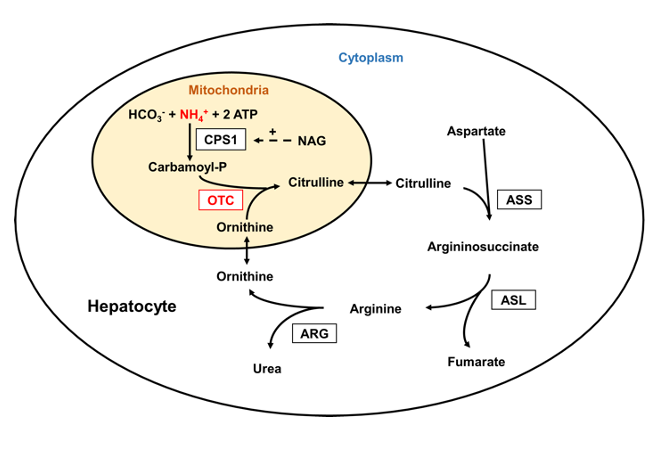
Source: Laemmle, Alexander; Gallagher, Renata C.; Keogh, Adrian; Stricker, Tamar; Gautschi, Matthias; Nuoffer, Jean-Marc; et al. (2016). Urea cycle.. PLOS ONE. Figure. https://doi.org/10.1371/journal.pone.0153358.g001
Glutamate, GABA and Sensitisation
Many patients describe symptoms that extend well beyond fatigue. These include:
Light, sound sensitivity
Sleep disturbance
Pain amplification
Cognitive overload
Anxiety-like physiological responses
Reduced tolerance of normal sensory input
One possible contributor is altered balance between two important neurotransmitters.
Glutamate is the brain's primary excitatory neurotransmitter
GABA is the brain's primary inhibitory neurotransmitter.
Together they help regulate neural activity. When glutamate activity becomes excessive relative to GABA, the nervous system may become increasingly reactive.
This process contributes to what is commonly known as central sensitisation, in which the nervous system becomes increasingly responsive to normal sensory, physical and cognitive inputs.
As inhibitory buffering declines, progressively smaller stimuli may produce disproportionately large symptom responses. Light, sound, movement, mental concentration or emotional stress may therefore trigger symptom amplification because the nervous system has lost part of its physiological reserve.
Many patients with severe PEM demonstrate amino acid profiles characterised by elevated glutamate and reduced GABA. While these findings do not establish causation, they suggest that altered neurotransmitter regulation may represent another important contributor to impaired recovery.
In this state, normal sensory, physical or cognitive demands can generate disproportionately large symptom responses. Many patients with severe PEM demonstrate amino acid profiles characterised by elevated glutamate and reduced GABA.
This does not prove causation. However, it suggests that neurotransmitter regulation may represent another important component of recovery failure.
The Missing Link: Pericytes and Oxygen Delivery
Energy production depends on oxygen. Without adequate oxygen delivery, mitochondria cannot function efficiently.
A specialised cell known as the pericyte plays a critical role in this process. Pericytes surround capillaries throughout the body.
They help regulate:
Capillary blood flow
Oxygen extraction
Blood–brain barrier integrity
Microvascular stability
Tissue perfusion
Under normal circumstances pericytes help match oxygen delivery to tissue demand.
Emerging evidence suggests that pericyte dysfunction may occur in Long COVID and other chronic inflammatory disorders. When pericytes become dysfunctional:
Capillary autoregulation may fail
Oxygen extraction becomes inefficient
Blood flow becomes mismatched to demand
Microregional hypoxia develops
Importantly, this can occur even when large blood vessels appear normal. The problem exists at the level of the microcirculation. This creates a critical link between vascular dysfunction and mitochondrial dysfunction.
Pericytes, the Kidney and Physiological Reserve
Pericytes are present throughout the body, including within the kidney where they help regulate microvascular blood flow, oxygen delivery and tissue stability.
The kidney plays a central role in sensing physiological stress and coordinating adaptive responses through the renin–angiotensin–aldosterone system (RAAS). Emerging evidence suggests that disturbances in renal microvascular function may influence how effectively the body responds to changes in posture, blood volume and physiological demand.
This possibility is of particular interest because many patients with Long COVID, POTS and related disorders demonstrate abnormalities of both recovery physiology and RAAS regulation. Ongoing research is exploring whether dysfunction of renal microvascular and pericyte-associated systems may contribute to impaired physiological reserve in some individuals.
While these observations remain preliminary, they suggest that recovery failure may involve interactions between the brain, microcirculation, metabolism and kidney rather than a single isolated biological pathway.
Figure 4. The Central Role of Pericyte–Neurovascular Dysfunction
This figure illustrates how early pericyte injury may disrupt capillary blood flow regulation, oxygen extraction and blood–brain barrier integrity. The resulting microregional hypoxia impairs mitochondrial function and activates chronic inflammatory pathways involving HIF-2α and STAT3. These changes create interconnected feedback loops linking hypoxia, neuroinflammation, metabolic dysfunction and impaired recovery.

Why Post-Exertional Malaise is Often Delayed
One of the defining features of PEM is that deterioration often occurs several hours after activity rather than immediately.
This delayed pattern suggests that symptoms do not simply reflect depletion of energy stores. Instead, the body initially compensates using its remaining physiological reserve. Heart rate, blood flow, autonomic regulation, oxygen delivery, mitochondrial metabolism and inflammatory control continue working to maintain function despite increasing physiological stress.
Eventually these compensatory systems become exhausted. As reserve declines, cerebral perfusion, oxygen delivery, mitochondrial energy production and neuroimmune regulation begin to deteriorate together. The result is delayed worsening of symptoms followed by prolonged recovery.
This helps explain why patients may initially feel capable of completing an activity before experiencing significant deterioration later that day or the following day.
When Recovery Failure Becomes Chronic
Acute physiological stress is normally reversible. However, when inflammatory signals, hypoxia or metabolic dysfunction persist, recovery pathways may begin to change.
Adaptive mechanisms designed for short-term survival become chronically activated.
Over time:
Oxygen delivery becomes less efficient
Mitochondrial performance declines
Inflammatory signalling persists
Tissue repair slows
Recovery capacity falls
Multiple biological systems begin communicating through self-reinforcing feedback loops. The result is a state where the illness becomes maintained by the interaction of several systems rather than by a single abnormality. This helps explain why recovery can be so difficult.
Patients are not dealing with one isolated problem. They are dealing with an interconnected biological network.
Persistent COVID Antibodies: What Do They Mean?
Many patients with Long COVID continue to demonstrate detectable or elevated SARS-CoV-2 antibody levels months or even years after the initial infection. This frequently raises the question of whether the virus remains active within the body.
At present, persistent antibodies should not be viewed as definitive evidence of ongoing viral replication. Long-lived plasma cells can normally produce antibodies for extended periods following infection, and several mechanisms may contribute to sustained antibody levels.
These include:
Long-lived immune memory
Persistent viral antigens or tissue reservoirs
Ongoing tissue injury and danger signalling
Failure of normal immune resolution pathways
Persistent activation of inflammatory signalling networks
Within the recovery-failure framework described in this paper, persistent antibody production may be viewed as a marker of ongoing immune activation rather than proof of active infection.
One pathway that may be particularly important involves chronic activation of the IL-6–STAT3 signalling axis. STAT3 is a key regulator of immune-cell survival, plasma-cell maintenance, inflammatory persistence and tissue remodelling. Persistent activation of STAT3 may therefore support continued antibody production while simultaneously contributing to chronic inflammation, hypoxia signalling and impaired physiological recovery.
In this model, elevated SARS-CoV-2 antibodies become part of a broader pattern of unresolved biological adaptation that may also include autonomic dysfunction, impaired oxygen delivery, neuroimmune activation, metabolic dysfunction and extracellular matrix remodelling.
Importantly, antibody persistence alone does not establish causation. Rather, it may serve as one indicator that recovery pathways have not fully returned to their pre-illness state.
In clinical practice, persistent antibody elevations are often observed alongside other markers of physiological dysregulation, including autonomic dysfunction, impaired exercise tolerance, neurocognitive symptoms and biochemical evidence of ongoing recovery failure. Whether antibodies are directly pathogenic or simply reflect an unresolved recovery state remains an active area of investigation.
Figure 5. From Acute Stress to Chronic Recovery Failure – A Pericyte-Centred Disease Model
This figure illustrates a proposed transition from acute physiological stress to persistent multi-system illness. Central to the model is endothelial–pericyte uncoupling, which promotes microvascular dysfunction, impaired oxygen delivery, chronic hypoxia, metabolic stress and neuroimmune activation. Multiple feedback loops subsequently develop involving inflammation, hypoxia, RAAS dysregulation, neurotransmitter imbalance and mitochondrial dysfunction, creating a self-sustaining state of recovery failure.

Disease Evolution: From Functional Dysregulation to Loss of Physiological Reserve
One of the most important observations emerging from Long COVID, POTS and ME/CFS research is that these conditions do not appear to remain biologically static over time.
In the early stages of illness, many abnormalities are predominantly functional. Pericyte dysfunction, impaired capillary autoregulation, intermittent hypoxia and autonomic instability may fluctuate substantially from day to day. Patients often describe periods of improvement followed by relapse, suggesting that physiological reserve remains present, even if impaired.
Over time, however, repeated cycles of hypoxia, inflammation and metabolic stress may begin to alter the tissues themselves.
When oxygen delivery repeatedly fails to match demand, hypoxia-inducible pathways become activated. Initially these responses are protective and help maintain short-term survival. However, prolonged activation may gradually shift cellular behaviour towards tissue adaptation rather than recovery.
Persistent activation of pathways involving RAGE, HIF-2α and STAT3 may promote:
Extracellular matrix remodelling
Fibroblast activation
Altered collagen organisation
Reduced tissue compliance
Impaired lymphatic function
Reduced venous buffering capacity
Loss of microvascular flexibility
As these structural adaptations accumulate, the body may become progressively less able to respond to physiological stress. This process can be viewed as a gradual loss of physiological reserve.
In practical terms, the body becomes increasingly constrained. Systems that once adapted easily to changes in posture, exercise, infection, emotional stress or environmental challenge become less flexible. Recovery takes longer. Symptom flares become easier to trigger and more difficult to reverse.
This framework may help explain an important clinical observation: patients with longstanding disease are often more difficult to treat than those in earlier stages of illness.
The problem may no longer be limited to transient dysregulation. Instead, years of repeated hypoxia, inflammation and metabolic stress may have produced measurable changes in tissue architecture, vascular behaviour and connective-tissue function.
Within this model, Long COVID evolves from a disorder of physiological dysregulation towards a disorder of altered tissue ecology.
The original trigger may have resolved long ago. However, the biological consequences of repeated microvascular stress continue to generate new danger signals, perpetuating activation of pathways such as RAGE, STAT3 and chronic hypoxia signalling. This creates self-sustaining loops in which tissue stress promotes inflammation, inflammation promotes remodelling, and remodelling further impairs physiological resilience.
Importantly, this concept does not imply irreversible disease. Rather, it suggests that treatment may become increasingly complex as illness duration increases, because both functional abnormalities and structural adaptations may need to be addressed simultaneously.
Each episode of incomplete recovery may leave slightly less reserve available for the next physiological challenge. Over time, this creates a downward spiral in which activities that were once easily tolerated progressively provoke larger and more prolonged symptom exacerbations. This gradual erosion of reserve provides a biological explanation for the progressive nature of many chronic illnesses.
When Compensation Creates New Problems
Long COVID is not simply persistent inflammation. It appears to be a progressive disorder of physiological reserve, driven by repeated cycles of pericyte dysfunction, regional hypoxia, metabolic adaptation and tissue remodelling.
One of the body's greatest strengths is its ability to adapt. However, prolonged compensation may create unintended consequences. As energy production becomes increasingly difficult, resources are progressively redirected towards immediate metabolic survival. This may leave fewer resources available for:
Connective tissue maintenance
Extracellular matrix repair
Collagen support
Vascular stability
Structural resilience
Over time, structural and metabolic dysfunction may begin reinforcing one another.
Reduced tissue integrity contributes to impaired perfusion
Impaired perfusion increases hypoxia.
Hypoxia further impairs mitochondrial function.
A self-perpetuating cycle develops.
Within this framework, the body remains biologically active rather than biologically inactive. Multiple adaptive systems continue attempting to restore normal function, but each cycle of compensation also consumes part of the remaining physiological reserve. Recovery therefore becomes progressively less efficient despite continued activation of protective pathways.
Figure 6. Metabolic–ECM Feed-Forward Loop Linking PDH Dysfunction to Structural Instability
This figure demonstrates how chronic metabolic dysfunction may gradually influence connective tissue integrity, vascular stability and biomechanical function. Reduced mitochondrial efficiency increases reliance on compensatory metabolic pathways, which may reduce resources available for tissue maintenance. The resulting structural instability may impair perfusion, increase hypoxia and further worsen metabolic dysfunction, creating a self-reinforcing biological loop.

“Adrenal Fatigue”
Many patients describe their illness using the term "adrenal fatigue" because they experience profound exhaustion, impaired stress tolerance, reduced resilience and an inability to recover normally following physical, cognitive or emotional demand.
While conventional endocrine testing often fails to demonstrate primary adrenal dysfunction, the symptoms themselves are real. The following figure illustrates a possible biological explanation. Rather than representing failure of the adrenal glands alone, these symptoms may arise from progressive loss of physiological reserve involving neurovascular, renal, autonomic, metabolic and stress-regulation systems.
Figure 7. From Physiological Stress to Recovery Failure: Loss of Physiological Reserve in Long COVID, POTS and ME/CFS
This figure illustrates how repeated physiological stress may progressively reduce the body's capacity to adapt and recover. Orthostatic stress, impaired haemodynamic adaptation, altered renin responsiveness, neurovascular dysfunction and endothelial–pericyte dysfunction progressively constrain multiple physiological reserve systems, including haemodynamic, neurovascular, metabolic, autonomic and inflammatory resolution pathways.
While patients may appear relatively stable at rest, increasingly minor physical, cognitive or emotional stressors can exceed the remaining reserve available for compensation. When this occurs, coordinated physiological regulation temporarily fails, producing post-exertional malaise (PEM). Rather than representing simple fatigue, PEM is proposed to reflect transient exhaustion of integrated physiological reserve.
Repeated episodes of incomplete recovery may progressively reduce reserve further, contributing to worsening fatigue, brain fog, autonomic dysfunction, impaired stress tolerance, sleep disturbance and increasingly prolonged recovery following exertion.
Within this framework, the syndrome commonly described as "adrenal fatigue" is more accurately interpreted as the clinical experience of impaired physiological reserve rather than primary adrenal dysfunction.

From this perspective, the syndrome commonly described as "adrenal fatigue" may represent the clinical experience of impaired physiological reserve rather than a disorder of adrenal hormone production itself.
Why Different Patients Look Different
Not all patients experience the same symptoms. Genetics, developmental factors, environmental exposures and physiological stressors influence how each person responds. Some patients develop predominantly autonomic symptoms. Others experience cognitive dysfunction. Some develop chronic pain or severe PEM.
Different pathways may dominate in different individuals.
However, many appear to converge on a common network involving:
Impaired oxygen delivery
Mitochondrial dysfunction
Neuroimmune activation
Metabolic compensation
Impaired recovery
Understanding this convergence helps explain why seemingly unrelated symptoms often occur together.
Different physiological reserve systems may become limiting in different patients. In some individuals, impaired haemodynamic reserve predominates, producing orthostatic intolerance and dizziness. Others demonstrate predominantly metabolic reserve failure, with severe post-exertional malaise. In others, neurovascular reserve or inflammatory resolution reserve may dominate the clinical picture. Although the symptoms differ, each reflects reduced capacity to maintain physiological homeostasis under stress.
Can Recovery Occur?
The existence of biological feedback loops does not imply permanence. It simply means recovery may require interruption of multiple reinforcing processes.
Different patients may require different approaches.
Some may benefit from improving blood flow
Others from addressing inflammation
Others from improving mitochondrial function.
Others from correcting mechanical, vascular or autonomic abnormalities.
The important concept is that recovery failure appears increasingly likely to represent a biological process rather than a failure of motivation, effort or resilience.
Understanding the mechanisms involved provides a framework for developing more effective treatment strategies and restoring physiological reserve.
Conclusion
Long COVID, POTS, ME/CFS and related disorders appear to involve far more than simple fatigue.
Emerging evidence suggests that impaired oxygen delivery, mitochondrial dysfunction, altered amino acid metabolism, neuroimmune activation and pericyte dysfunction may interact to create a state of persistent recovery failure.
Within this framework, symptoms such as fatigue, PEM, brain fog, sensory hypersensitivity and autonomic dysfunction can be understood as different manifestations of an interconnected biological network.
Long COVID, POTS, ME/CFS and related disorders appear to involve far more than simple fatigue. Emerging evidence suggests that impaired oxygen delivery, mitochondrial dysfunction, altered amino acid metabolism, neuroimmune activation, endothelial–pericyte dysfunction and impaired physiological resolution interact to create a state of persistent recovery failure.
Within this framework, symptoms such as fatigue, post-exertional malaise, brain fog, sensory hypersensitivity and autonomic dysfunction can be understood as different manifestations of progressive loss of physiological reserve rather than isolated diseases affecting individual organs.
Understanding these illnesses through the concept of physiological reserve provides a practical framework for explaining why recovery becomes increasingly difficult over time. Rather than representing irreversible organ damage, these conditions appear increasingly to reflect failure of multiple adaptive systems to recover fully following physiological stress.
Identifying the dominant processes limiting physiological reserve in each individual may ultimately provide the most rational path towards more personalised, mechanism-based approaches to recovery.


Comments